Introduction
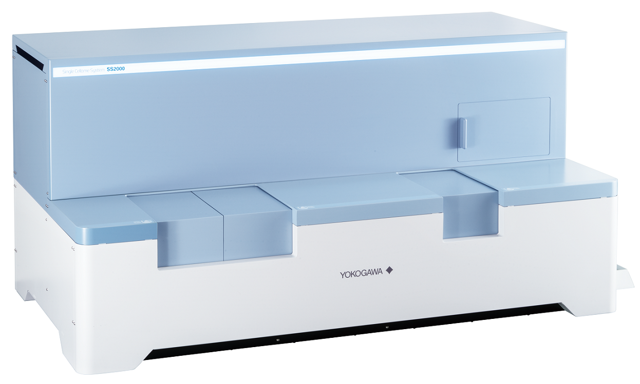
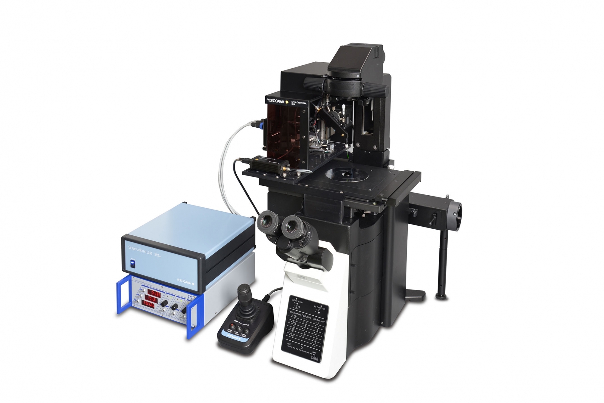
The SS2000 is a system that automatically samples specific regions of cells or whole cells at the single-cell level while imaging cells in culture with a confocal microscope. Because cells in culture do not need to be detached, positional and morphological information is maintained.
Collected samples can be transferred to PCR plates and microwell plates. It is also possible to accumulate multiple samples into the same well or retain it in the glass tip for removal without ejecting. The sample collection site is equipped with a cooling function to prevent sample degradation and has an incubating function to maintain culture conditions. Collected samples can be used for genetic analysis, mass spectrometry, single-cell cloning, and more.
It is based on the technology of the live cell imaging product developed by our company. High-speed, high-resolution 3D imaging is possible using our unique confocal microscope technology. Samples can be taken from targeted cells under a confocal microscope in an incubator environment. Time-lapse photography is also possible, allowing dynamic changes in the target cell to be captured. Since it is possible to record moving images during sampling and images before and after sampling, it is possible to compare the results of analysis of collected samples with cell imaging data.
Features / strengths
1. Special Glass Tips
- Inner diameter of tips 3μm 5μm 8μm 10μm
- One rack contains 96 tips
2. Automatic target selection
- Target cells and sampling positions can be automatically selected by imaging analysis. (Targets can be automatically selected as shape of cells, size of nuclei, density of organelles etc.)
3. Tile imaging & Illumination uniform tool
- Tile imaging can efficiently capture images of the entire wide field of view. Illumination uniform tool Uniformizer is installed as standard equipment, and image seams can be captured uniformly
4. Incubator function
- maintaining cell activity
Specification in detail
Tip diameter
3μm 5μm 8μm 10μm
Incubator loader environment
37°C, 5%CO2, humidified
Excitation laser wavelength
405, 488, 561, 640 nm
Objective lens
Dry lens: 4x, 10x, 20x, 40x Long-working distance lens: 20x, 40x Note that only the 40x dry lens can be used for cell sampling.
Camera
sCMOS camera 2,000 x 2,000 pixel Pixel size: 6.5 x 6.5μm
Operating environment
Temperature: 15 to 30°C Humidity: 30 to 70%RH no condensation
Data formats (Measurement software)
Captured images: 16bit TIFF (OME- TIFF, TIFF) Output image data: TIFF, PNG, JPEG Output video data: WMV, MPEG4
Data formats (Analysis software)
Numeric data: CSV Output image data: TIFF, PNG, JPEG Output video data: WMV, MPEG4
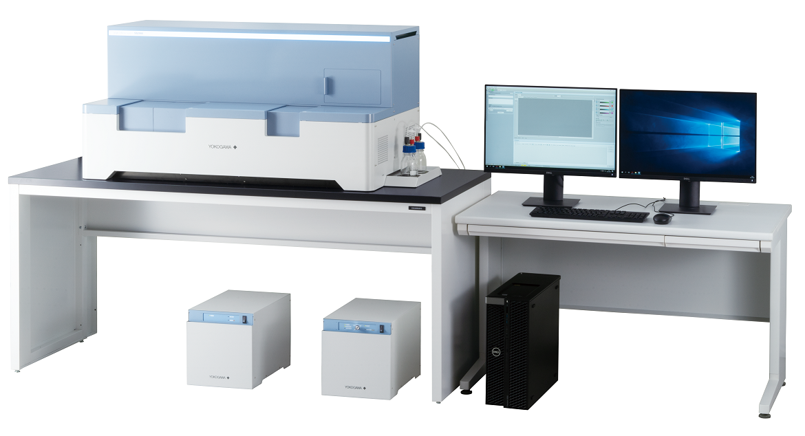
External dimensions , Weight
Main unit: W1,217 x D643 x H595 mm/145kg Utility box: W275 x D432 x H298 mm/18kg Gas mixer: W275 x D432 x H298 mm/10kg Special purpose workstation: W172 × D471 × H414 mm/14kg Display: W531 x D500 x H166 mm/5.6kg
Supporting document