Introduction
Within the drug development market, demands on high content analysis systems for drug efficacy evaluation are increasing in accordance with the needs for cell-based assay and phenotypic screening. In order to increase screening efficiency, devices with higher speeds (higher throughput) are required. On the other hand, in order to bridge the “valley of death” of the drug development process, the quality of screening hits must be increased. This requires the construction of more complex evaluation systems that utilize multifaceted parameters via 3D cultivation systems, live-cell imaging and higher detail image analysis. In current drug development research, determining how to implement throughput screening and complex valuation system screening in parallel is an important issue.
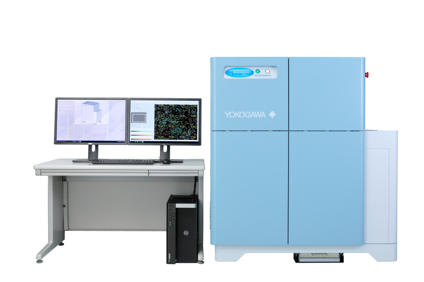
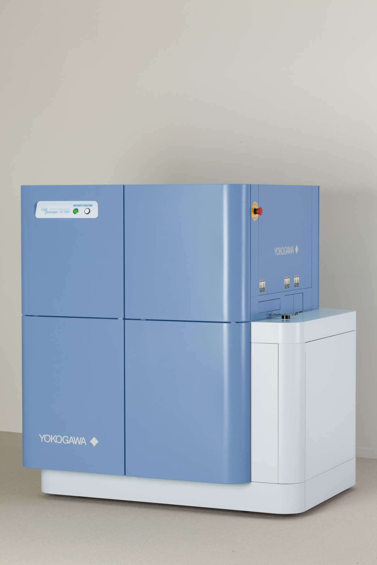
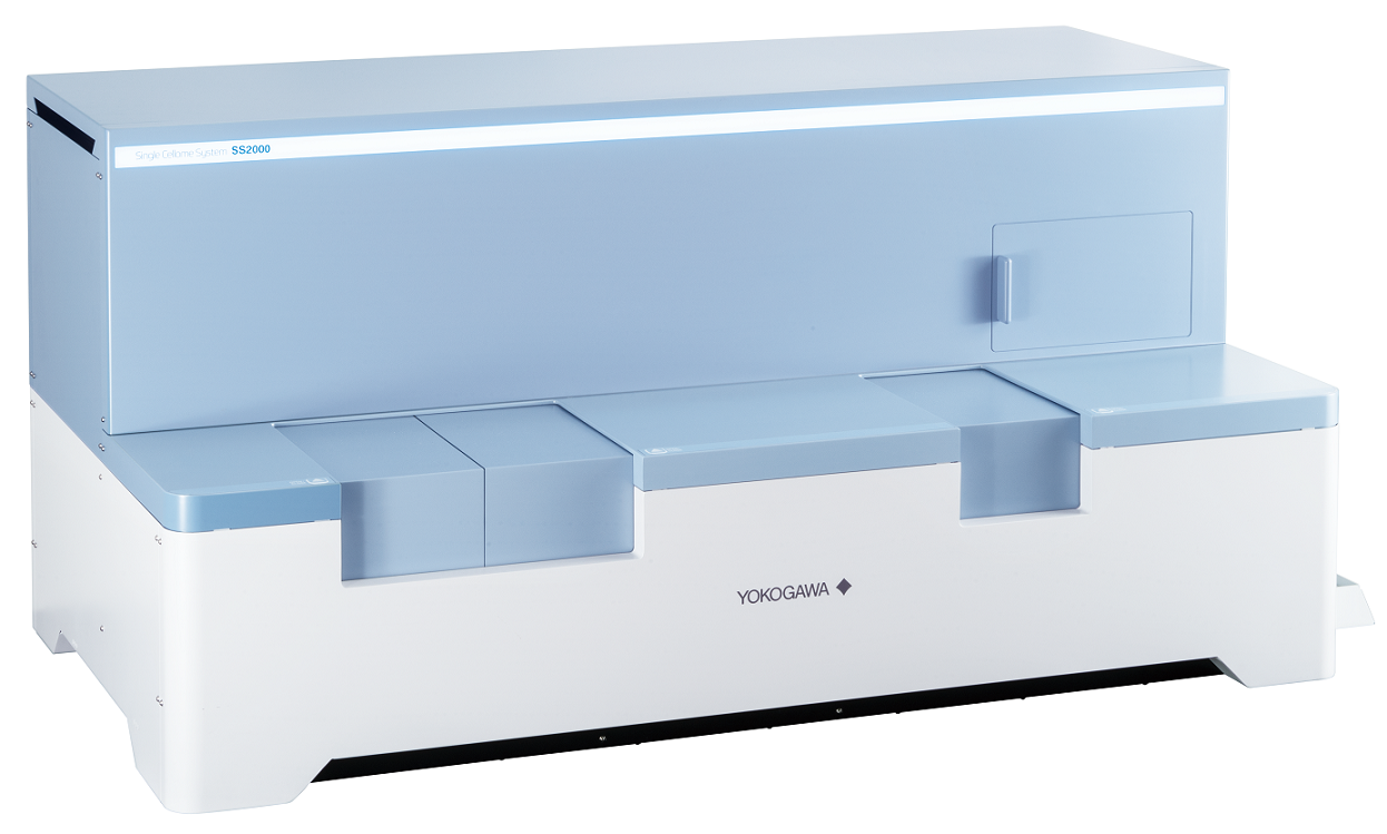
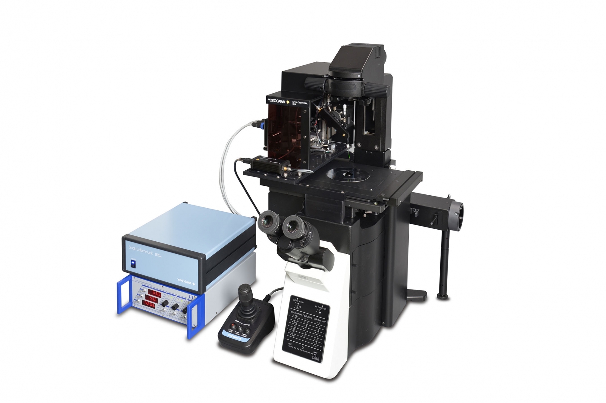
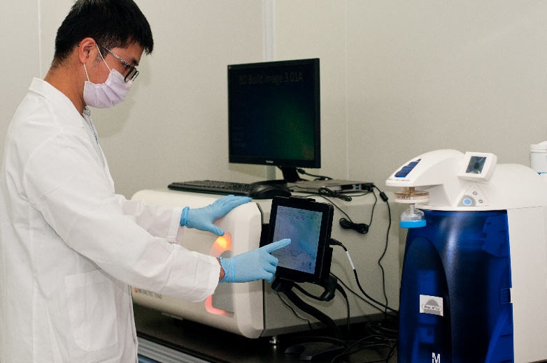
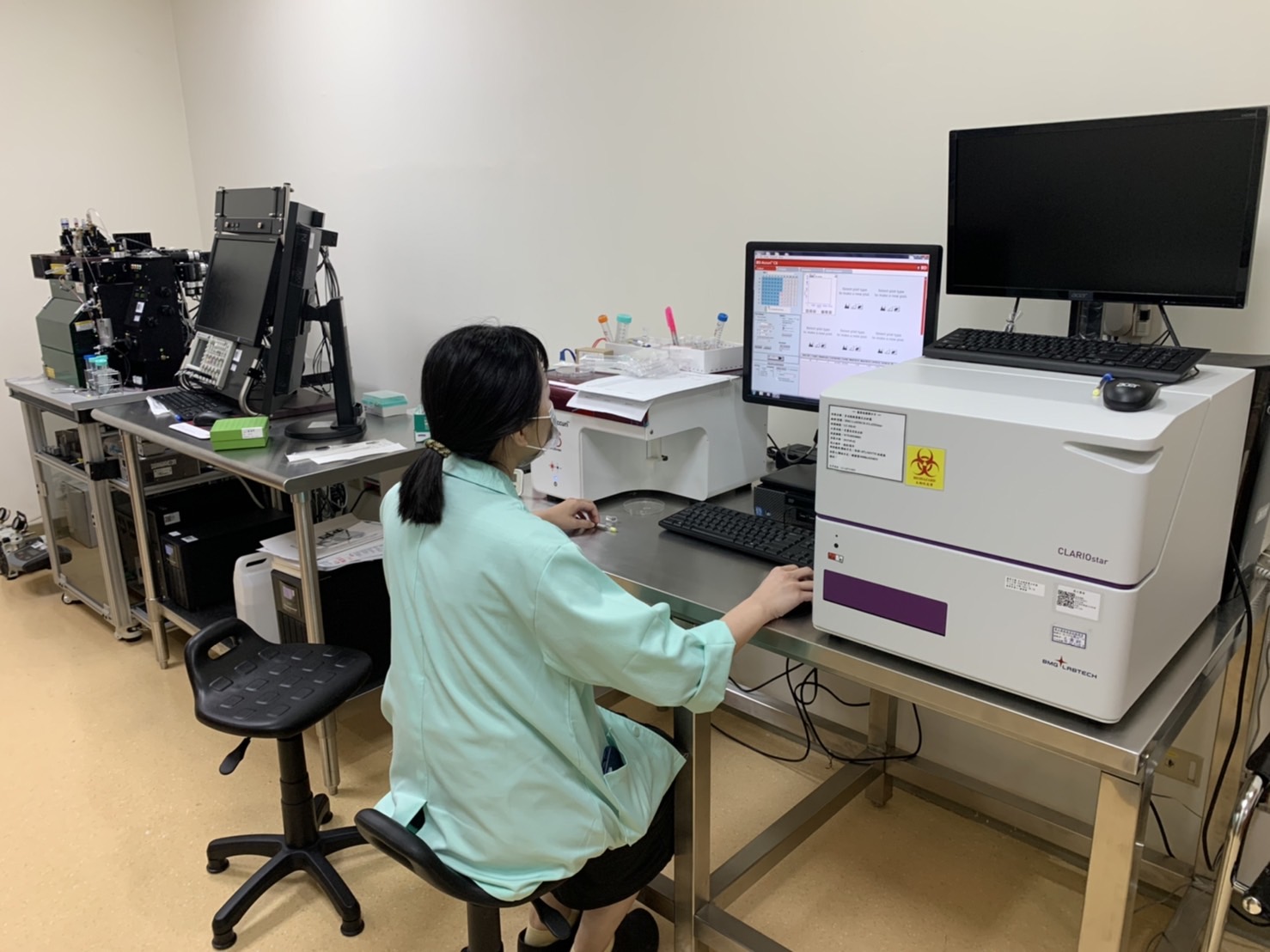
CV8000 is a high-end, high content analysis system that solves this contradictory screening challenge. Through the combination of a proprietary Company high speed confocal scanner, water immersion lens, up to four high field-of-vision cameras, a microscopic stage with cell cultivation environment, and an integrated robotic pipetter, we have realized not only high throughput, high-resolution imaging, but also phenotypic screening via a more complex evaluation system. In addition, our specialized analysis software, Software, uses deep leaning and machine learning to recognize target objects with high accuracy, supporting you from image analysis to results display using graphs.
Features / strengths
Long-term livecell imaging
- Stage incubator included as standard. Realization of non-stop, long-duration observation (3 days +) via humidity, temperature and CO2 control.
Kinetic assay
- Drug addition during imaging is made possible by an integrated robotic pipetter with disposable tips.
- Ideal for kinetic experiments involving the observation of high speed phenomena.
Organoid / Spheroid
- Yokogawa’ s spinning disk confocal technology excels in imaging of samples with depth, such as 3D culture samples where clear and quick imaging is difficult, allowing for evaluation close to in-vivo quality.
Label-free analysis
- Recognition and analysis can be performed by taking bright field images from several Z positions and creating a CE bright field image using the included CellPathfinder analysis software. Analysis accuracy is further enhanced via the new Deep Learning option.
Specification in detail
Sample format
Multiple well plate (6, 12, 24, 48, 96, 384, 1536 wells), glass slide
Output data format
Image data: 16bit TIFF, PNG Numerical data: CSV, original format
Excitation wavelength
405/445/488/561/640 nm, all solid laser, max. 5 lasers 【Option】365 nm LED
Confocal unit
Microlens-enhanced wide-view dual Nipkow disk confocal scanner, 50 μm pinhole 【Option】 25 μm pinhole disk and auto pinhole disk exchanger
Camera
sCMOS (effective pixels: 2000X2000 pixel size: 6.5 μm), max. 4 cameras
Stage incubator
Temperature for incubation : 35-40℃ CO2 supply box (CO2: 5%, forced humidification)
Operating environment
15~30℃ 30~70%RH (no condensation)
Dimensions
W1,280×D895×H1,450 mm;510Kg
Supporting document