Introduction
Clear 3D images obtained from confocal microscopes have been enabling advancements in cell biology research for many years.
This imaging technology combined with population analysis now provides a significant advancement for cytometry.
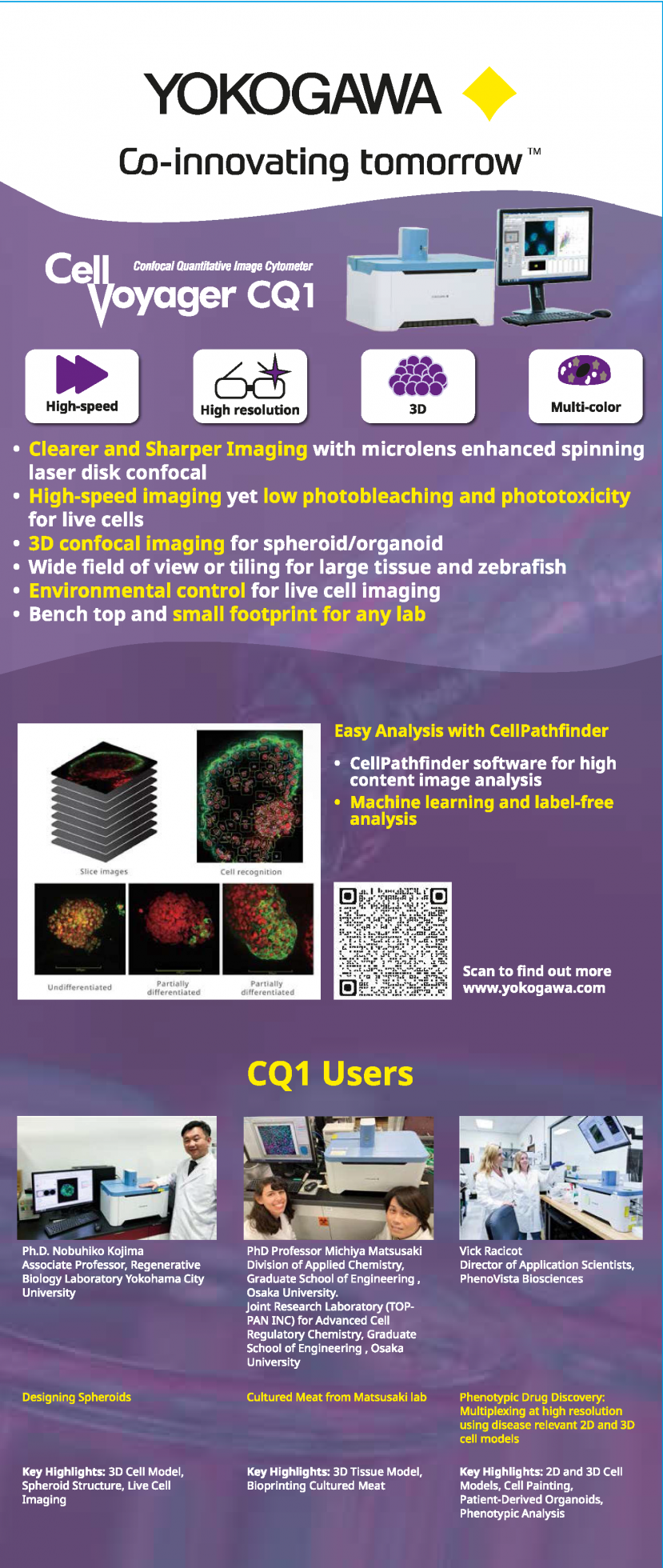
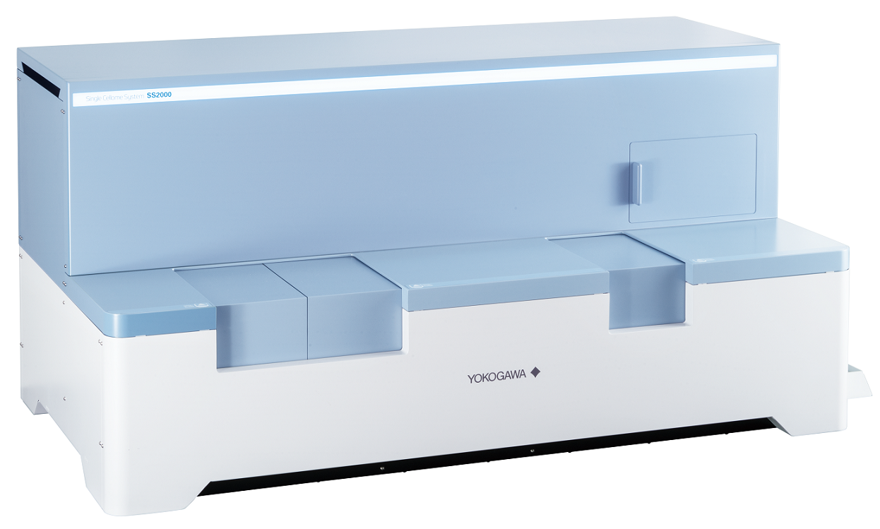
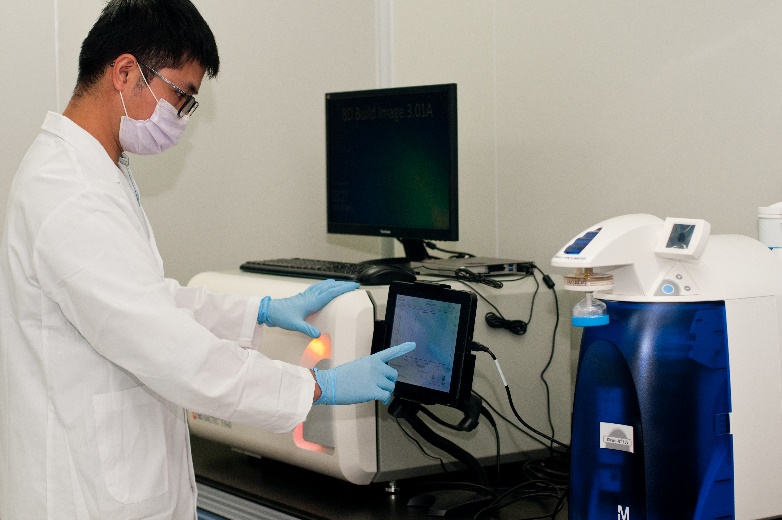
The CQ1 enables clear 3D imaging, object recognition, and rapid quantification of live cells and cell clusters.
The data from the images help in the understanding, and enhance the reliability of data.
The CQ1's live cell chamber acts as a cellular incubator enabling is used to many times lapse imaging while the CQ1's unique
imaging technology is gentle on the cells.
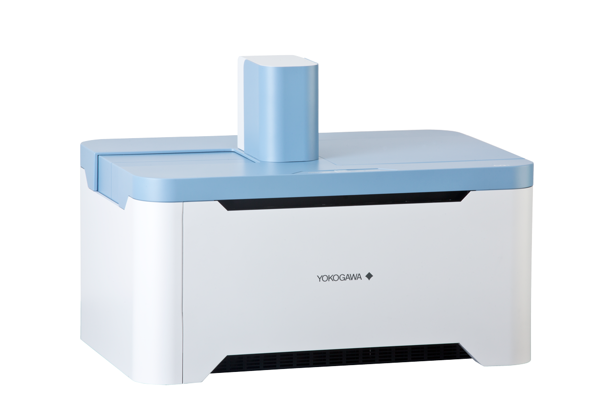
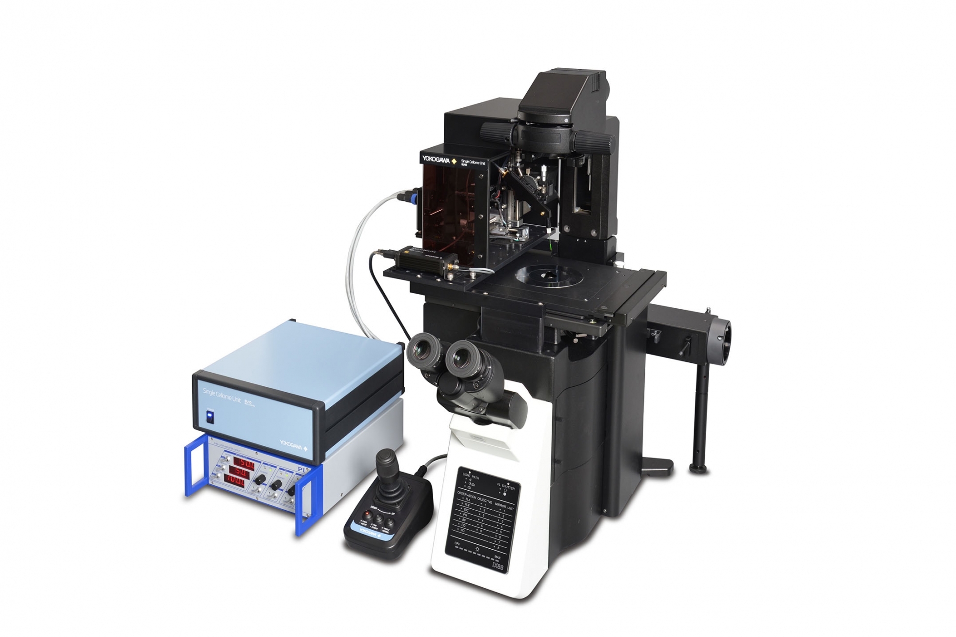
The Yokogawa CQ1 is an easy to use all-in-one confocal microscope for a reasonable price.
The CQ1 comes with a number of configurable options and can be integrated into a fully automated screening system.
Features / strengths
1. Enables measurement of spheroids, colonies, and tissue sections
No need to remove cells from the culture dish, in contrast to traditional flow cytometry
Nipkow spinning disk confocal technology allows high-speed yet gentle 3D image acquisition
Rich feature extraction to facilitate sophisticated cellular image analysis
Wide field of view and tiling capability enables easy imaging of large specimen
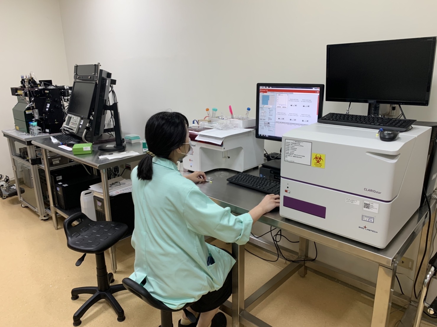
2. Enables analysis of time-lapse and live-cell
High precision stage incubator and low phototoxicity of our confocal makes the analysis of time-lapse and live-cell are possible
Max. 20fps option for fast time-lapse
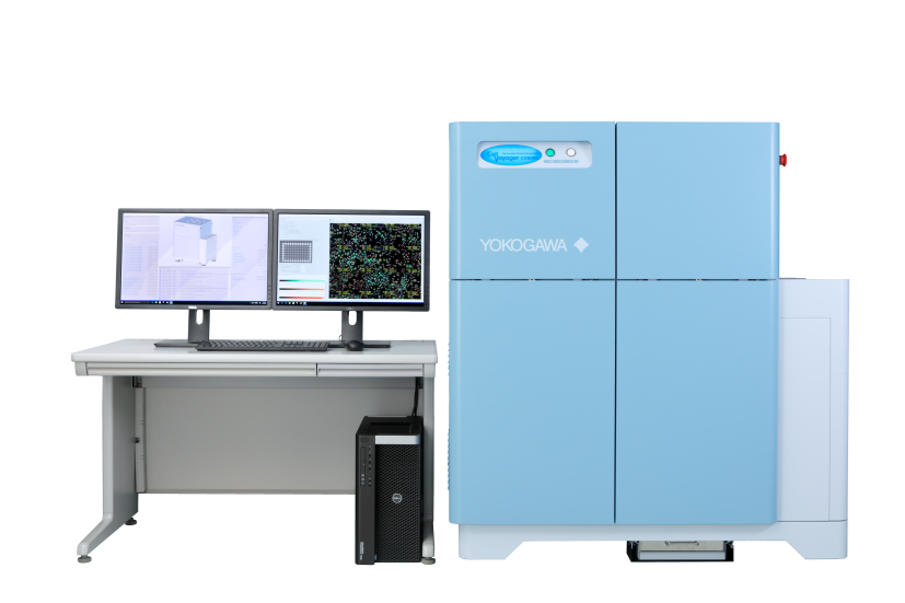
3. High-quality image and similar operability to a traditional flow cytometer
Integration with CellPathfiner software can provide powerful analysis displayed in real-time with image acquisition
Usable high-quality image as confocal microscope image.
Interactive graphs make it easy to trace back the data points
4. Open platform
Connectable with external systems via handling robot*2
Expandable to the integrated system as image acquisition and quantification instrument
FCS/CSV/ICE data format readable by third-party data analysis software
A variety of cell culture and sample dishes are applicable
Specification in detail
Fluorescence
Laser : Choose Max.4 lasers from 405 / 488 / 561 / 640 nm EM Filter : Max. 10 filters (Included 1 filter for transmitted illumination)
Objective lens
Max.6 lenses Dry : 2x, 4x, 10x, 20x, 40x Long working distance : 20x, 40x For thick bottom vessel : 20x Phase contrast*1 : 10x, 20x
Data format
Captured image : 16 bit TIFF (OME-TIFF) Output image format : TIFF (16 bit, 8 bit) , PNG, JPEG Output movie format : WMV, MP4 Output numerical data format : FCS, CSV, ICE
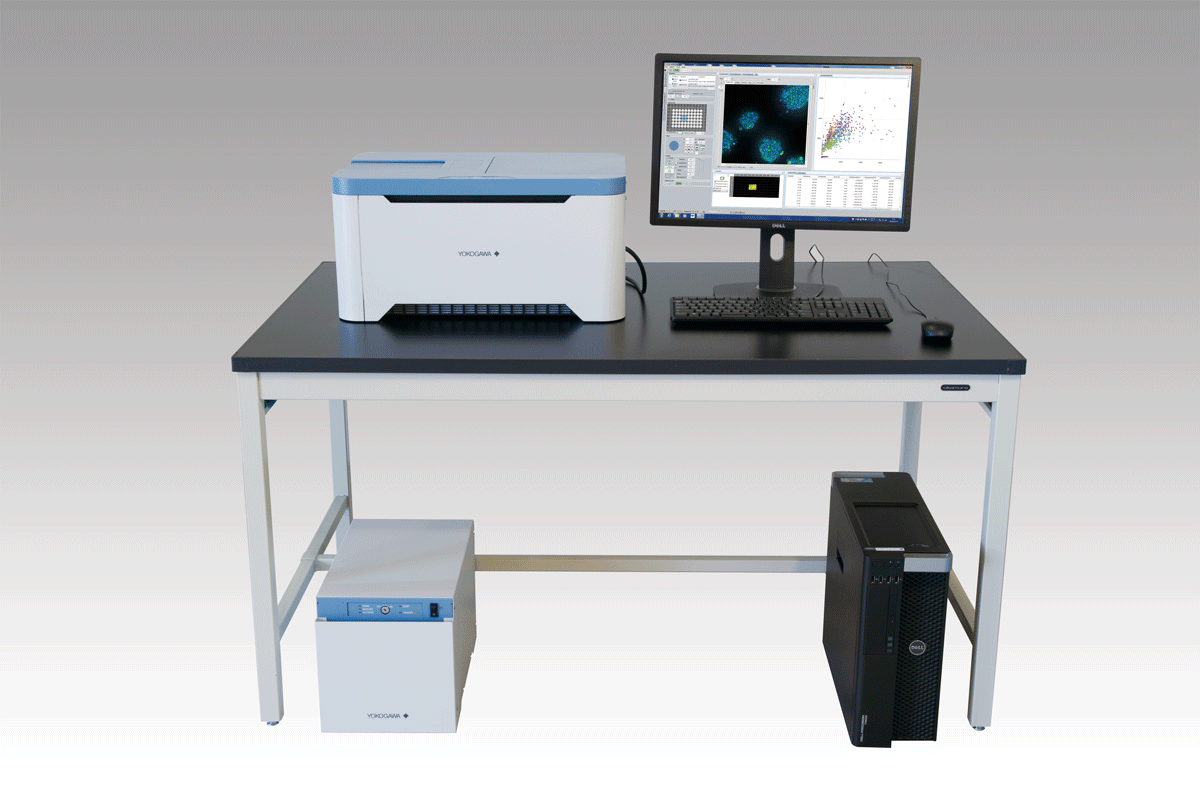
Size / Weight
Main unit : 600 × 400 × 437 mm, 44 kg Utility box : 275 × 432 × 298 mm, 18 kg Gas Mixer (Option) : 170 × 260 × 280 mm, 5.2 kg
Environment
Main unit and Utility box : 15 – 35 °C, 20 – 70 % RH No condensation Gas Mixer (Option) : 20 – 30 °C, 10 – 85 % RH No condensation
Power consumption
Main unit and Utility box : 100-240 VAC, 800 VAmax Workstation : 100-240 VAC, 950 VAmax Gas Mixer (Option) : 100-240 VAC, 50 VAmax
Supporting document