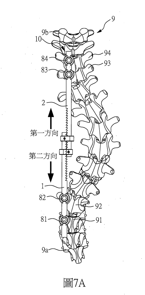
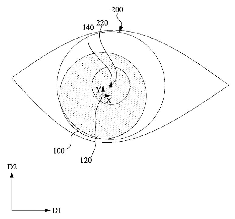
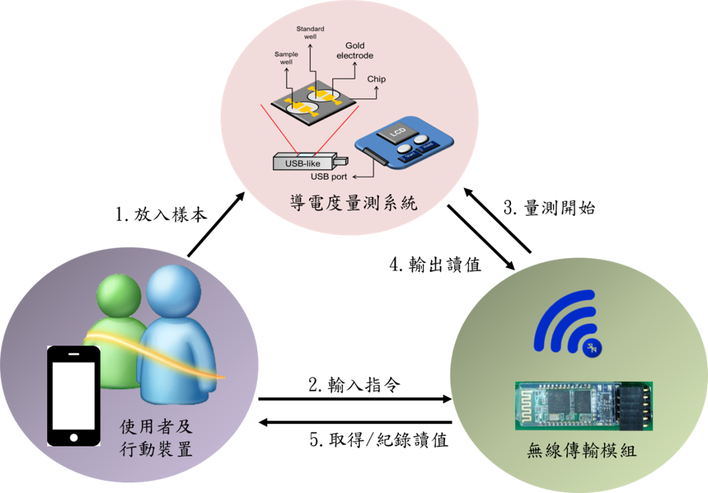
Introduction
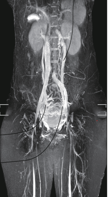
This patented technology is a non-invasive medical imaging examination that does not require using a contrast medium or producing a cumulative radiation dose. It is highly suitable for women, children, patients with chronic kidney disease, and those with adverse reactions to contrast medium. This technology can aid in diagnosing pelvic venous diseases (such as pelvic congestion syndrome) and lower extremity venous diseases. It can meet various clinical needs and be utilized for developing related medical applications. The technology employs two-dimensional scanning to obtain images of pelvic and lower extremity venous structures and uses three-dimensional scanning to assess the location of vascular anomalies. In addition, this technology quantitatively analyzes whether blood flow changes are consistent with clinical symptoms through cross-sectional blood flow measurements. Compared to ultrasound examinations, this patented technology is superior in detecting pelvic venous diseases and performs better in identifying deep blood vessels in the lower limbs.
Features / strengths
This patented technology is a non-invasive medical imaging examination that does not require using a contrast medium or producing a cumulative radiation dose. It is highly suitable for women, children, patients with chronic kidney disease, and those with adverse reactions to contrast medium. This technology can aid in diagnosing pelvic venous diseases (such as pelvic congestion syndrome) and lower extremity venous diseases. It can meet various clinical needs and be utilized for developing related medical applications. The technology employs two-dimensional scanning to obtain images of pelvic and lower extremity venous structures and uses three-dimensional scanning to assess the location of vascular anomalies. In addition, this technology quantitatively analyzes whether blood flow changes are consistent with clinical symptoms through cross-sectional blood flow measurements. Compared to ultrasound examinations, this patented technology is superior in detecting pelvic venous diseases and performs better in identifying deep blood vessels in the lower limbs.
Specification in detail
Intellectual Property Office, MOEA
I815656